The Retina — Your Eye’s Most Powerful Processor
Have you ever wondered how your eyes convert a glance at a colourful sunrise into a vivid, three-dimensional image in your brain? The answer lies in a paper-thin sheet of tissue at the back of your eye — the retina. And within that tissue, there are not one but 10 distinct layers, each performing a unique and essential job.
Understanding the 10 layers of the retina is not just for medical textbooks. It is the foundation for appreciating why conditions like diabetic retinopathy, age-related macular degeneration (AMD), and glaucoma can be so devastating — and why early detection saves sight.
According to the National Institutes of Health (NIH), the retina is composed of six different cell lines organized into ten functional layers. Together, these layers make the retina the most metabolically active tissue in the entire human body — consuming more oxygen per gram than even the brain.
In this article, we walk you through each of the 10 layers of the retina in order, explain their individual functions, and show you how damage to any single layer can affect your vision. Whether you are a student, a patient, or simply curious, this guide will help you see your own eyes in a whole new light.
What Is the Retina? A Quick Overview
The retina is a thin, light-sensitive tissue lining the inner back wall of the eye. Despite being less than half a millimetre thick, it contains millions of specialized nerve cells organized into a precisely layered architecture.
Think of it like the sensor in a digital camera. When light enters your eye and passes through the lens, it is focused onto the retina. The retina captures that light, converts it into electrical signals, and transmits those signals to the brain via the optic nerve. The brain then interprets these signals as the images we perceive.
The key to all of this is the retina’s layered structure. Each of the 10 layers of the retina has a specific role in this process — from absorbing stray light and nourishing photoreceptor cells to processing visual information and transmitting nerve signals.
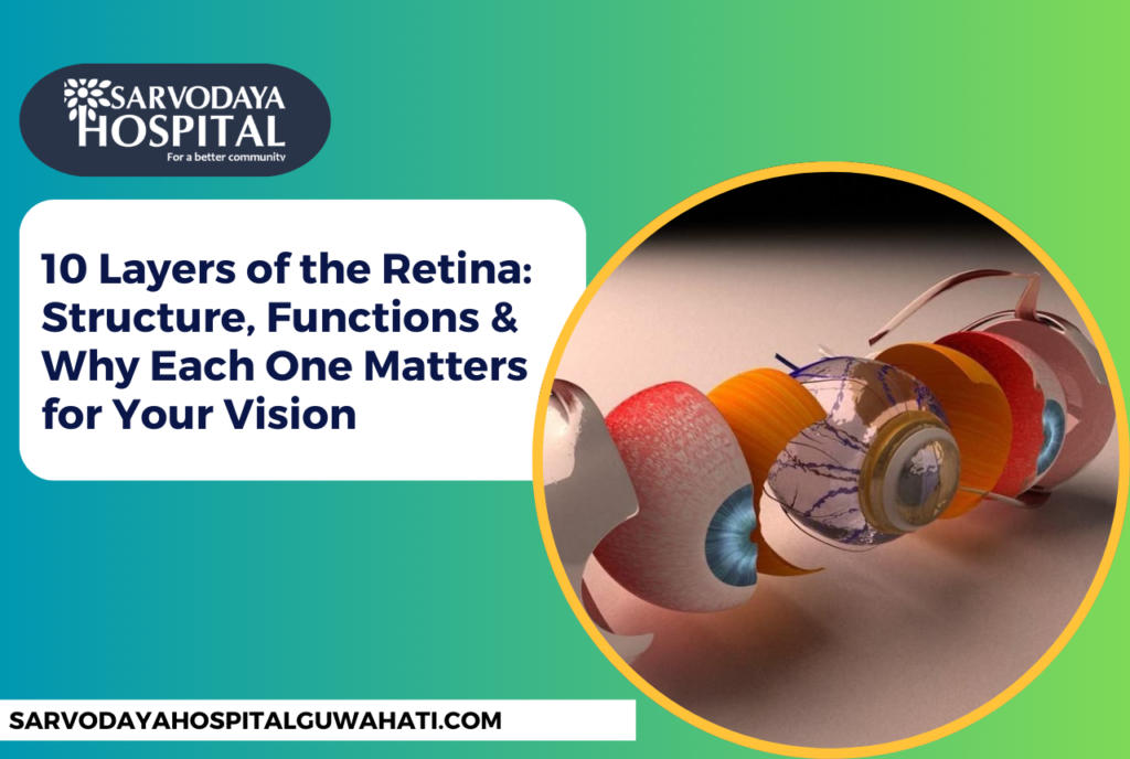
The 10 Layers of the Retina: In Order from Outside to Inside
According to Wikipedia’s detailed retinal anatomy reference, the 10 layers are arranged from the outermost position (closest to the choroid, the eye’s blood vessel layer) to the innermost position (facing the vitreous humour, the gel filling the eye).
Layer 1: Retinal Pigment Epithelium (RPE)
The RPE is a single layer of hexagonal, pigmented cells sitting at the very outer edge of the retina. It is the unsung hero of eye health.
Functions: The RPE absorbs scattered light to prevent glare and blurring, transports vital nutrients and oxygen from the choroid to the photoreceptors, removes metabolic waste products from the outer retina, and recycles visual pigments (such as Vitamin A/retinal) that are essential for phototransduction.
Why it matters: Dysfunction of the RPE is the primary driver of Age-Related Macular Degeneration (AMD), the leading cause of irreversible blindness in people over 60 worldwide.
Layer 2: Photoreceptor Layer (Rods & Cones)
This is where vision truly begins. The photoreceptor layer contains the outer and inner segments of two types of photoreceptor cells.
- Rods: Around 120 million rods provide vision in low-light (scotopic) conditions and are essential for peripheral and night vision.
- Cones: Approximately 6–7 million cones are concentrated in the central macula and are responsible for sharp colour vision and fine detail (photopic vision) in bright light.
When light strikes the photopigments inside rods and cones, it triggers a chemical reaction called phototransduction — converting light energy into an electrical signal. This is the foundational step of all vision.
Layer 3: Outer Limiting Membrane (OLM)
The OLM is not a true membrane in the traditional sense but a series of junction complexes formed by Müller cells — the major support cells of the retina. It acts as a structural boundary between the photoreceptor inner segments and the outer nuclear layer. It controls the exchange of molecules, maintaining the integrity and organization of the photoreceptors.
Layer 4: Outer Nuclear Layer (ONL)
The ONL contains the cell bodies (nuclei) of the rod and cone photoreceptors. While the photoreceptor layer holds the active tips of these cells that capture light, the ONL is where their genetic and metabolic machinery lives. The thickness of the ONL is a reliable indicator of photoreceptor health and can be measured using Optical Coherence Tomography (OCT).
Layer 5: Outer Plexiform Layer (OPL)
The OPL is a synaptic zone — a communication hub. Here, the axon terminals of the photoreceptors (called rod spherules and cone pedicles) connect with the dendrites of bipolar cells and horizontal cells. These connections form the first synapses in the visual pathway, where raw light data begins to be processed and refined. This is where signals begin their journey toward the brain.
Layer 6: Inner Nuclear Layer (INL)
Often called the processing centre of the retina, the INL is densely packed with the cell bodies of three key neuron types:
- Bipolar Cells: Transmit visual signals from photoreceptors toward the ganglion cells.
- Horizontal Cells: Adjust signal strength between neighbouring photoreceptors, helping the eye adapt to both bright and dim environments (lateral inhibition).
- Amacrine Cells: Over 30 subtypes are involved in complex processing tasks such as motion detection, edge enhancement, and contrast modulation.
Layer 7: Inner Plexiform Layer (IPL)
The IPL is another critical synaptic zone — perhaps the most complex in the retina. As explained by the StatPearls/NIH Neuroanatomy of the Retina, the IPL functions like a switchboard where bipolar cell axons connect with the dendrites of ganglion cells and amacrine cells. The IPL is subdivided into five sub-layers, each carrying a different type of elementary visual information — particularly the separation of “ON” pathways (responding to light increase) and “OFF” pathways (responding to light decrease).
Layer 8: Ganglion Cell Layer (GCL)
The GCL contains the cell bodies of retinal ganglion cells (RGCs) — the retina’s final output neurons. There are approximately 20 different types of RGCs, each specializing in different aspects of visual information such as motion, colour, contrast, and spatial frequency.
Notably, 1–2% of all RGCs are intrinsically photosensitive (ipRGCs) and contain a photopigment called melanopsin. These cells are not involved in conscious vision but instead regulate circadian rhythms, pupil size, and melatonin release — making them central to sleep and overall health.
Layer 9: Nerve Fiber Layer (NFL)
The NFL consists of the long axons of ganglion cells that sweep across the inner retinal surface, converging at the optic disc (the blind spot) to form the optic nerve. These axons carry all processed visual information from the retina to the brain’s visual cortex.
Clinical importance: The NFL is critically important in glaucoma diagnosis. Elevated eye pressure in glaucoma damages these nerve fibers, and thinning of the NFL detected by OCT scanning is one of the earliest signs of the disease — often before any noticeable vision loss occurs.
Layer 10: Inner Limiting Membrane (ILM)
The ILM forms the innermost boundary of the retina, composed of the basement membrane of Müller cells. It acts as an interface between the retina and the vitreous humour — the gel-like substance that fills the main chamber of the eye.
The ILM provides structural support to the retina and helps maintain its shape and position. In vitreoretinal surgery, surgeons carefully peel the ILM to treat conditions such as macular holes and epiretinal membranes (internal scar tissue).
Common Retinal Disorders Linked to These Layers
Now that you understand the 10 layers of the retina, it becomes clear why various eye diseases can be so vision-threatening. Here is a brief overview (you can find more information at the Discovery Eye Foundation):
- Age-Related Macular Degeneration (AMD): Primarily affects the RPE and photoreceptor layers, leading to central vision loss.
- Diabetic Retinopathy: Damages the blood vessels supplying the inner retinal layers, causing fluid leakage, swelling, and eventually haemorrhage.
- Retinal Detachment: Physically separates the photoreceptor layer from the RPE, cutting off the nutrient supply and causing rapid, permanent vision loss if untreated.
- Glaucoma: Progressive damage to the NFL and GCL, leading to irreversible peripheral and central vision loss.
- Retinitis Pigmentosa: A genetic condition causing progressive degeneration of rod photoreceptors, beginning with night blindness.
What Are the Early Signs of Retinal Damage?
Retinal disease can be painless and silent in its early stages. Key warning signs to watch for include:
- Sudden increase in floaters or flashes of light
- A dark shadow or curtain across part of your visual field
- Blurred or distorted central vision (straight lines appearing wavy)
- Difficulty seeing in low light or at night
- Loss of peripheral (side) vision
If you experience any of these symptoms, it is important to seek prompt evaluation from an eye specialist. At Sarvodaya Hospital Guwahati, our ophthalmology team uses advanced OCT (Optical Coherence Tomography) imaging to visualize all 10 layers of the retina in real time and detect problems at their earliest, most treatable stage.
Can the Retina Repair Itself?
Unlike some tissues in the body, the mature retina has very limited capacity for self-repair. Photoreceptor cells, once lost, do not regenerate in humans. This is why prevention and early intervention are so critically important.
However, active research in stem cell therapy and gene therapy is showing enormous promise. Conditions like Leber Congenital Amaurosis (LCA) have already been treated with gene therapy in clinical trials, with patients experiencing meaningful improvements in vision. The unique immune-privileged environment of the eye makes it an ideal candidate for such treatments.
For the latest developments in retinal research, visit the National Eye Institute (NEI).
How to Protect Your Retinal Health Naturally
While you cannot fully regenerate a damaged retina, you can take meaningful steps to protect the health of all 10 layers:
- Eat a diet rich in antioxidants, particularly lutein and zeaxanthin (found in leafy greens, eggs, and corn), which are concentrated in the macula.
- Wear UV-protective sunglasses to reduce oxidative damage to the RPE.
- Manage systemic conditions like diabetes and hypertension, which directly threaten retinal blood supply.
- Avoid smoking, which dramatically increases the risk of AMD and reduces blood flow to retinal layers.
- Stay physically active — regular exercise has been shown to improve ocular blood flow.
- Schedule regular comprehensive eye examinations, especially if you are over 40 or have a family history of retinal disease.
Looking for the Best Retina Specialist in Guwahati?
If you live in Guwahati or anywhere in Northeast India and are concerned about your retinal health, getting the right specialist matters. The retina’s 10 intricate layers require advanced diagnostic tools and deep clinical expertise to assess accurately — and not every eye clinic is equipped for that.
At Sarvodaya Hospital Guwahati, our ophthalmology department is equipped with Optical Coherence Tomography (OCT) — the gold standard in retinal imaging — which allows our specialists to scan and examine every individual layer of your retina with extraordinary precision. This makes it possible to detect early-stage conditions like macular degeneration, diabetic retinopathy, and glaucoma-related NFL thinning before symptoms even begin.
When searching for the best retina specialist in Guwahati, look for these key qualities:
- Formal subspecialty training in vitreoretinal surgery or medical retina
- Access to advanced diagnostic equipment including OCT, Fundus Fluorescein Angiography (FFA), and wide-field retinal imaging
- Experience managing both medical retinal conditions (AMD, diabetic retinopathy) and surgical cases (retinal detachment, macular holes)
- A patient-centred approach with clear communication and personalized treatment plans
Our team at Sarvodaya Hospital Guwahati is trusted by thousands of patients across Assam and the wider Northeast region. Whether you need a routine retinal screening or a specialist consultation for a complex condition, we are here to help you preserve and protect your most precious sense — your sight.
Conclusion: See the Bigger Picture
The 10 layers of the retina represent one of nature’s most elegant engineering achievements. In a tissue no thicker than a single human hair, millions of specialized cells are organized into 10 distinct strata, each contributing to the remarkable gift of sight. From the RPE that quietly nourishes photoreceptors to the ILM that forms the retina’s innermost boundary, each layer is indispensable.
Understanding these layers is not merely an academic exercise — it is the clinical foundation for diagnosing and treating the eye diseases that affect millions of people globally. Modern imaging tools like OCT allow retinal specialists to examine each layer individually, enabling earlier diagnosis and better outcomes than ever before.
At Sarvodaya Hospital Guwahati, we are committed to bringing world-class retinal care to our patients. If you have concerns about your vision or retinal health, do not wait. Book a consultation with our eye specialists today.